SPECIMEN STABILITY CONSIDERATIONS DURING FLOW CYTOMETRY ASSAY DEVELOPMENT, OPTIMIZATION AND VALIDATION

- Flow Cytometry User Groups And The Relaunch Of The NWFCS - August 31, 2023
- Fc Receptors (FcR) and their Relevance to Flow Cytometry - July 5, 2022
- Announcing the addition of Cytek® Aurora technology to our available instrumentation - January 25, 2022
Introduction
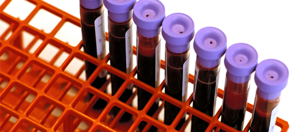
Flow cytometry assays are widely used to evaluate Pharmacodynamic (PD) effects of molecules during all phases of drug development. Specimens for flow cytometry analysis are collected throughout the course of a study and often shipped to a central location for testing. Testing at a central location mitigates variability resulting from differences in instrumentation, reagents, and analysis methodology. Typically samples are shipped the day they are drawn resulting in a processing and analysis delay of 1-3 days for fresh whole blood or specimens can be stored frozen and tested in batches. As a result, specimen stability must be determined as part of method development and implementation.
Stability Evaluation Process Overview
The assessment of specimen stability begins with a process to determine stability based on the intended assay performance. Specimen stability must be carefully evaluated over an appropriate time course starting from specimen collection through sample acquisition. Each stability parameter requires evaluation during method development and validation. If the specimen stability does not meet the requirements for the intended use of the assay, re-configuring the assay may result in increased stability.Stability of a specimen can be affected by the following variables:
- Downstream Assay Requirements
- Specimen Type
- Specimen Collection Methods
- Specimen Storage
- Shipping Conditions
This process can be divided into two parts. Initially the focus will be on the stability of the specimen during blood draw or specimen collection. The emphasis of these stability considerations is on determining the assay objectives, the type of specimen collected and anticoagulant required. Next, after the specimen is collected, the process for assessing specimen stability is based on assay performance. This includes defining the specimen stability window, setting acceptance criteria and determining the impact, if any, of the storage and shipping conditions.

Figure 1: A flow chart illustrating a process for determining stability in flow cytometry specimens. It is recommended that the assay parameters be chosen first and then the stability is determined by the assay performance. If specimen stability does not meet the assay performance then the assay parameters may need to be re-evaluated.
Stability Assessment Parameters
Peripheral whole blood is the most frequently used specimen collected for flow cytometry analysis. However, some assays require preparation and freezing peripheral blood mononuclear cells (PBMC) rather than whole blood. Tissue specimens such as bone marrow, spleen, thymus, lymph node and tumor can also be collected for flow cytometry analysis. These tissues must be disrupted into single cell suspensions prior to analysis. If solid tissue specimens will be shipped then the logistics of when, where and how the cell suspensions are to be prepared should be evaluated during assay development, if possible.Whole blood and bone marrow can be collected into blood collection tubes containing an anticoagulant or in some cases a preservative. The choice of anticoagulant is driven by both the specimen stability and the type of flow cytometry assay utilized (i.e., phenotyping, antigen expression, or functional assays). In general, Sodium Heparin and EDTA are versatile anticoagulants for multiple applications in the laboratory. These can be considered acceptable anticoagulants for flow cytometry unless data exists to suggest the use of a different anticoagulant for a specific application. If extended stability is required, cell stabilization products such as CytoChex BCT that contain both an anticoagulant and a cell preservative may allow for longer periods of stability.
Since specimens collected during preclinical and clinical studies are usually shipped to a central lab for analysis; it is prudent to evaluate the effect of shipping conditions on these specimens. The specimens can be packaged in a shipping container with temperature buffering agents such as ambient or refrigerated gel packs. The package can either be stored on the bench top or transported to mimic transit. If specimen temperature is critical to specimen stability for a particular flow cytometry assay, then temperature tracking should be considered. There are a variety of commercially available monitoring options that are available to record package temperatures during shipping.
Several factors should be considered when establishing stability acceptance criteria. Precision of the assay is often used as guidance for establishing specimen stability acceptance criteria. Relative percent change is also a common descriptive statistic used to measure stability. This is the percent change between the fresh specimen and the stored specimen. However, stability assessments may also include a careful examination of assay analysis plots and/or histograms for changes in assay results. Light scatter properties as well as surface marker stability may be altered, depending on the temperature the specimens are exposed to prior to processing. For example, the granulocyte population of a whole blood specimen collected in EDTA and Sodium Heparin is a sensitive cell population and may show degradation of forward and side scatter within 24 hours of collection, when held at room temperature.
With the understanding that no single solution will be applicable to every scenario possible, this article proposes a standardized process for assessment of specimen stability for use with a variety of flow cytometry assays.
For more information please sign up for our guides:
Shipping and Stability Guide for Biological Specimens Part 1: Stability Considerations during Assay Development and Optimization
Shipping and Stability Guide for Biological Specimens Part 2: Stability Assessment Planning, Data Evaluation and Acceptance Criteria
References:
Recommendations for the evaluation of specimen stability for flow cytometric testing during drug development. Authors: Lynette Brown, Cherie L Green, Nicholas Jones, Jennifer J Stewart, Stephanie Fraser, Kathy Howell, Yuanxin Xu, Carla G Hill, Christopher A Wiwi, Wendy I White, Peter J O’Brien, Virginia Litwin. J Immunol Methods 2015 Mar 4;418:1-8. Epub 2015 Feb 4.