EMT6 Tumor
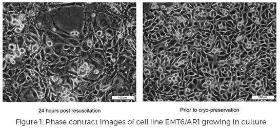
EMT6 is an epithelial tumor of the mammary glands of BALB/cCrgl Mus musculus. This tumor has produced an adherent cell that requires storage in liquid nitrogen. EMT6 has been used in mice to study the antitumor potential of Cutibacterium (Propionibacterium) acnes, the bacteria responsible for acne in humans, and melatonin. Metformin and curcumin’s ability to keep breast cancer from proliferating has been tested utilizing this tumor cell line. The EMT6 tumor has also been used to verify the effect of CD200 on tumor growth.
A study published in 2013 focused on curing metastatic growth of EMT6 in BALB/c mice by manipulating the signals of CD200:CD200R. Previously CD200 was thought to contribute to cancer progression by an increase in expression of the molecule. Immunocompromised mice expressing CD200, either via the host or tumor cells, lead to an increase in seeding of the tumor cells to the draining lymph node, as opposed to their immunocompetent counterparts. When CD200 was neutralized by anti-CD200mAbs the tumor metastasis and cytotoxic anti-tumor immune cells decreased in the draining lymph node. After excision, the mice remained tumor free for up to a year.
At FCSL we have performed immunophenotyping on many tumor types, looking at a variety of lymphocyte subsets. Stayed tuned for more information and our poster presentation this fall at PANWAT, taking place this year October 14-15 at Seattle Genetics, Inc. in Bothell, Washington which will dive into this tumor type plus a whole lot more!
References
“ECACC General Cell Collection: EMT6/AR1.” Public Health England, European Collection of Authenticated Cell Cultures, www.phe-culturecollections.org.uk/products/celllines/generalcell/detail.jsp?refId=96042327.
“EMT6/P CB_96042344.” Sigma-Aldrich, The Journal of Pharmacy and Pharmacology, 2018, www.sigmaaldrich.com/catalog/product/sigma/cb_96042344?lang=en.
Gorczynski, Reginald M., et al. “Cure of Metastatic Growth of EMT6 Tumor Cells in Mice Following Manipulation of CD200:CD200R Signaling.” Breast Cancer Research and Treatment, vol. 142, no. 2, 2013, pp. 271–282., doi:10.1007/s10549-013-2735-3.