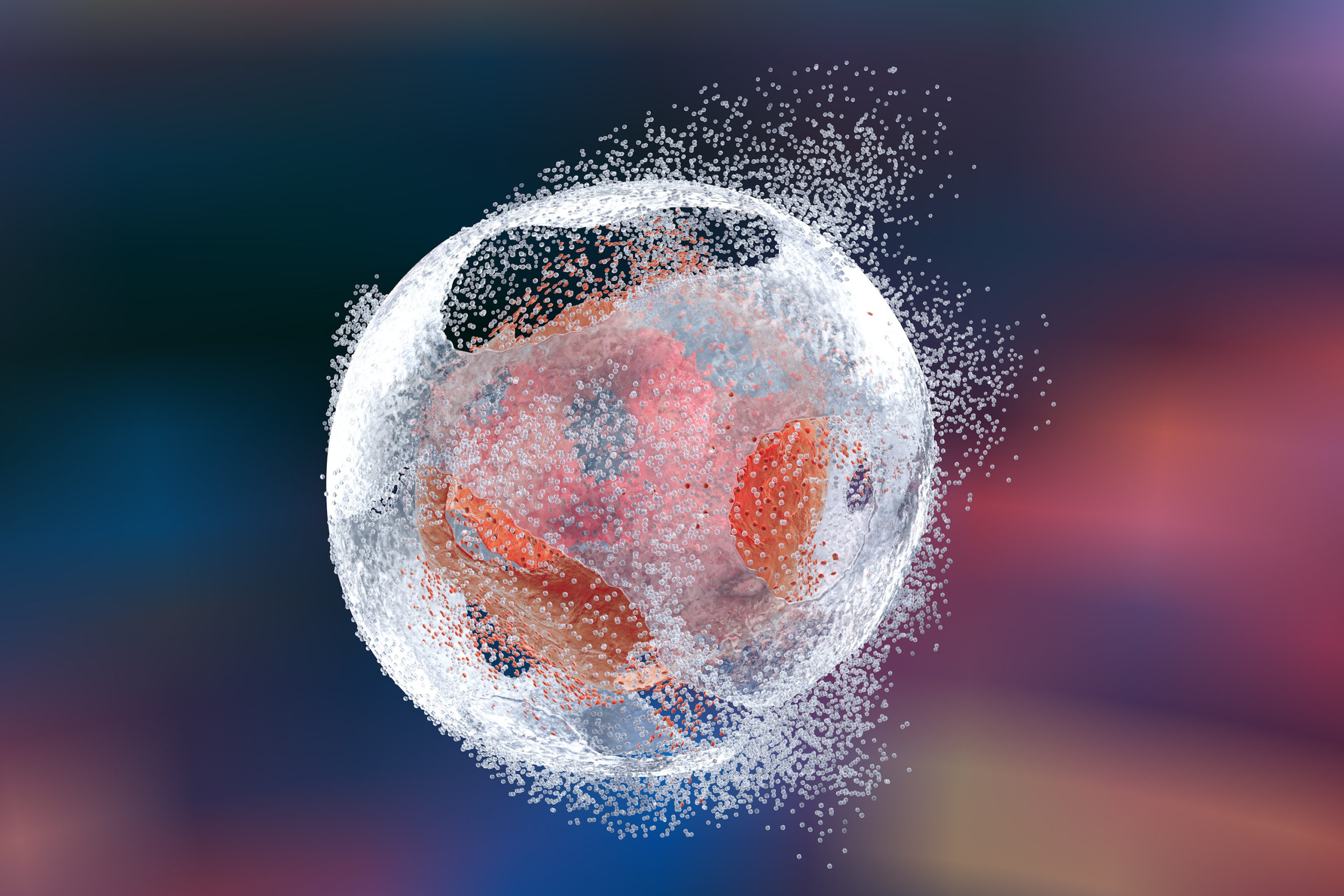
Live Dead Measurement and Cell Viability
- Live Dead Measurement and Cell Viability - August 29, 2022
Most antibodies cannot cross the intact cell membrane of a live cell and thus antibodies attach themselves to the proteins bearing their target antigen present on the outer membrane. However, as cells die, their cell membrane is disrupted and becomes permeable. This can cause fluorophore labeled antibodies to bind specifically and nonspecifically to the dead cell (1,5). Due to this, dead cells take up antibodies and can be mistaken as positive events during Flow Cytometry analysis without live cell discrepancy. The first step to differentiate between the live and dead cells is to use light scatter as dead cells have distinctive properties (Figure 1). This is done by plotting forward scatter area against forward scatter height to remove any doublets, as dying cells often clump to each other. However, light scatter is not dependable enough to use by itself (5). The next step is to use viability dyes, which is essential to gate out the remainder of the dead and dying cells. These dyes will stain the dead cells allowing the analyst to target viable cells during analysis.
In functional assessments such as antibody-dependent cell-mediated cytotoxicity (ADCC), these types of assays use viability dyes like, Calcien AM and Celltrace, that cross the cell membrane of live cells and are detected by hydrolysis of esterase in the cytosol. These methods allow for the positive staining of live cells instead of dead cells.
Viability dyes that stain dead cells are broken into two main types, fixable and non-fixable dyes. Non-fixable dead cell stains refer to DNA binding dyes. DNA binding dyes will cross the permeable membrane of the dead cells and bind to DNA. The DNA binding dyes will not bind to the DNA of live cells because the cell membrane is still intact. Dead cells will then fluoresce, or become positive, and live cells will remain negative. Thus, allowing for distinction of viable cells during analysis.

7-Aminoactinomycin D (7-AAD) and Propidium Iodide (PI) are two common DNA binding dyes used in flow cytometry. 7-AAD binds to double stranded DNA by intercalating in G-C base pair areas. Propidium iodide (PI) binds to double stranded DNA by intercalating base pairs with little or no sequence preference. However, (Rieger et al., 2010) shows that PI staining results in a large percentage, up to 40%, of false positive events when processed in conventional staining methods (4). Other DNA binding dyes include, Sytox® which binds to both DNA and RNA and DaPi (4′,6-diamidino-2-phenylindole) which binds to double stranded DNA by intercalating in T-A base pairs areas (6).
DNA binding dyes are not always favorable for use in intracellular processes because the fixation and permeabilization can compromise intact cell membranes. However, non-fixable dyes can be used for intracellular staining assays, but only if added prior to the fixation and permeabilization steps. When DNA binding dyes are added after cells are fixed and permeabilized the dyes cross the membrane of live cells and bind with the DNA, creating a falsely positive stained sample (Figure 3). Instead, fixable dead cell stains (amine-reactive dyes) are often used in an intracellular process when cells will be fixed and permeabilized. LIVE/DEAD™ Aqua, Zombie Green™, and LIVE/DEAD™ NIR are three different amine-reactive dyes. These “fixable” dyes cross the membrane of dead cells then react with free amines in the cytoplasm (2,3). Live cells will not bind with the amine-reactive dyes due to their intact membranes. when the dye is added, the leftover dye that does not bind with the dead cells free amines will be washed away before the fix and permeabilize steps. When the cells are fixed the dye stays within the dead cells, this is considered an irreversible process (2).
Viability dyes are a vital part of Flow Cytometry in order to differentiate between the dead and live cells. Zombie dyes and LIVE/DEAD™ Dyes are brands of amine reactive dyes available through BioLegend® and Invitrogen™ that we have featured above. They come in many excitation and emission wavelengths which can allow for a diverse and adaptable antibody panel design (3).
FCSL is a contract flow cytometry lab with expertise in complex flow cytometry panel & assay design, including Receptor Occupancy Assays (ROA), Immunophenotyping, Cell Signaling, Cell Cycle Analysis, and Cell Functional Assays. Often these types of flow cytometry assays require discrimination between live and dead cells for the most robust results. Here at FCS Laboratory, we have an incredible team to help figure out which viability dye will be best in your assay.
References
- O’Brien, M. C., & Bolton, W. E. (1995). Comparison of cell viability probes compatible with fixation and permeabilization for combined surface and intracellular staining in flow cytometry. Cytometry, 19(3), 243–255.
- Perfetto, S. P., Chattopadhyay, P. K., Lamoreaux, L., Nguyen, R., Ambrozak, D., Koup, R. A., & Roederer, M. (2010). Amine-reactive dyes for dead cell discrimination in fixed samples. Current Protocols in Cytometry, Chapter 9, Unit-9.34.
- Perfetto, S. P., Chattopadhyay, P. K., Lamoreaux, L., Nguyen, R., Ambrozak, D., Koup, R. A., & Roederer, M. (2006). Amine reactive dyes: An effective tool to discriminate live and dead cells in polychromatic flow cytometry. Journal of Immunological Methods, 313(1), 199–208.
- Rieger, A. M., Hall, B. E., Luong, L. T., Schang, L. M., & Barreda, D. R. (2010). Conventional apoptosis assays using propidium iodide generate a significant number of false positives that prevent accurate assessment of cell death. Journal of Immunological Methods, 358(1), 81–92.
- Schmid, I., Hausner, M. A., Cole, S. W., Uittenbogaart, C. H., Giorgi, J. v, & Jamieson, B. D. (2001). Simultaneous flow cytometric measurement of viability and lymphocyte subset proliferation. Journal of Immunological Methods, 247(1), 175–186.
- Estandarte, A. K., Botchway, S., Lynch, C., Yusuf, M., & Robinson, I. (2016). The use of DAPI fluorescence lifetime imaging for investigating chromatin condensation in human chromosomes. Scientific Reports, 6(1), 31417. https://doi.org/10.1038/srep31417