To Fc or not Fc: An assay for the question

- To Fc or not Fc: An assay for the question - August 1, 2022
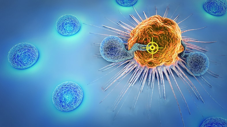
Our immune systems are in a constant battle with the thousands of pathogens that we encounter every day. Immune cells have established versatile tactics to defend against intruders. One mechanism in particular has intrigued researchers due to the invaluable implications it retains. Antibody-dependent cell-mediated cytotoxicity (ADCC) is a mechanism utilized by the immune system as a defense strategy against target cells expressing tumor or pathogen derived surface antigens. In this process, the surface of the target is coated by specific antibodies with exposed FC regions that bind FC receptors bearing cells called effector cells. Effector cells include natural killer cells, eosinophils, mast cells and activated T cells (1) . The binding of the FC receptor and the FC region acts as a bridge between the effector cell and the target allowing for direct delivery of toxic contents such as perforin/granzymes and cytotoxic granules (2). As a result, the target is fated to destruction (Figure 1). This effective defense strategy can be mimicked in vitro to develop assays to study cell mediated responses. For example, Doepker et al. 2021 inquired about the elicitation of desirable antibodies pertaining to vaccine development and this assay was used to screen antibodies that effectively mediate ADCC (3).

Figure 1. FAB regions of the antibodies bind to the antigen of the target cell with their FC regions exposed. The FC receptors found on effector cells bind to the FC region of the antibody. This binding triggers the release of toxic molecules directly to the target cell (1A). The toxic molecules result in the lysing of the target cell (1B).
Throughout the years, different methods for ADCC quantification have been available. Previously, 51Cr release assay has been widely used to quantify cytotoxicity. This method is reproducible and simple to perform however, low sensitivity and poor labeling of target cell lines have been reported. Disposal of radioactive substances poses major biohazardous challenges (4,5). Live cell imaging through fluoresce microscopy has also been used to quantify ADCC however some flaws have been documented. Shortcomings include limited available parameters and exposure time to fluorescent light thus compromising the experiment to maintain health of cells therefore limiting the amount of time the experiment can be conducted(6).
Quantification of the ADCC assay is best interpreted through flow cytometry. The high-throughput equipment allows for highly reproducible in-depth analysis at the single cell level of multiple samples in a cost and time efficient manner. Furthermore, analysis of this assay through flow cytometry allows phenotypic classification of effector through the identification of subset populations within a sample due to multiparameter capabilities. Therefore, the flow cytometer can simultaneously sort out live and dead cells allowing for the quantification of destroyed target cells. ADCC assay analysis through flow cytometry presents low biohazardous risks, allows for more samples thus producing more data, in a reasonable time span and is therefore best suited for generating meaningful results from the ADCC assay
A straightforward approach to evaluate and rank the ability of antibodies to induce ADCC in response to antigen detection is to label target cells with fluorescence such as GFP. The antibodies of interest are incubated with the target cells. Next the addition of effector cells, in a known ratio to target cells, are added. These cells are incubated, and the samples are then stained with viability dyes such as 7-actinomycin D dye (7-AAD) to distinguish nonviable cells when acquired on the flow cytometer. Additional antibody staining may be performed to detect certain effector cells. By gating methods shown below and simple computations, the percentage of dead target cells can be used to compare the strengths amongst different antibodies (Figure 2). Comparisons amongst activated effector cells may also be statistically determined and illustrated by similar gating methods. Furthermore, the same experiment can be done to evaluate ADCC response by antibodies in a dose-dependent manner.


FCSL is a contract flow cytometry lab that routinely runs standard and custom flow cytometry panels supporting basic research, non-clinical and clinical studies. We are experts in fit-for-purpose flow cytometry assays, immunophenotyping of lymphocyte subsets, receptor occupancy assays (ROA) as well as development of cellular functional assays such as the ADCC assay. We also offer services such as cell culturing and frozen PBMC processing; both of which may be necessary for complete execution various functional assays. If your research would benefit from immune monitoring using the ADCC assay, contact us to find out how our flow cytometry services can support your program.
References
- Dhanji S, Tse K, Teh H-S. The Low Affinity Fc Receptor for IgG Functions as an Effective Cytolytic Receptor for Self-Specific CD8 T Cells. The Journal of Immunology 2005;174:1253–1258.
- Gómez Román VR, Murray JC, Weiner LM. Antibody-Dependent Cellular Cytotoxicity (ADCC). Antibody Fc: Linking Adaptive and Innate Immunity 2014:1–27.
- Doepker LE, Danon S, Harkins E, Ralph D, Yaffe Z, Garrett ME, Dhar A, Wagner C, Stumpf MM, Arenz D, Williams JA, Jaoko W, Mandaliya K, Lee KK, Matsen FA, Overbaugh JM. Development of antibody-dependent cell cytotoxicity function in HIV-1 antibodies. Elife 2021;10:1–45.
- Zahavi D, AlDeghaither D, O’Connell A, Weiner LM. Enhancing antibody-dependent cell-mediated cytotoxicity: A strategy for improving antibody-based immunotherapy. Antibody Therapeutics 2018;1:7–12.
- Yamashita M, Kitano S, Aikawa H, Kuchiba A, Hayashi M, Yamamoto N, Tamura K, Hamada A. A novel method for evaluating antibody-dependent cell-mediated cytotoxicity by flowcytometry using cryopreserved human peripheral blood mononuclear cells. Scientific Reports 2016;6.
- Robson AL, Dastoor PC, Flynn J, Palmer W, Martin A, Smith DW, Woldu A, Hua S. Advantages and limitations of current imaging techniques for characterizing liposome morphology. Frontiers in Pharmacology 2018;9.