Silent but Deadly, and Beneficial? – Natural Killer Cells

- The Technology Behind Spectral Flow Cytometry - April 22, 2022
- Silent but Deadly, and Beneficial? – Natural Killer Cells - November 21, 2018
- Comparison of Different Fix/Permeabilization Buffers for use in Flow Cytometry Evaluations - August 22, 2018
Silent but Deadly, and Beneficial? – Natural Killer Cells
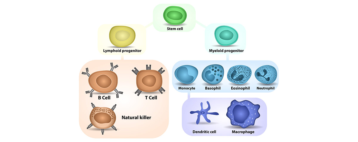
Natural Killer (NK) cells play a major role in targeting and killing both tumor cells and virally infected cells in the body. Although they play a very similar role to cytotoxic T cells, these cells comprise an important component of the host’s innate immune system. Most immune cells of the adaptive immune system detect the antigens presented by the major histocompatibility complex (MHC) on the surface of infected cells, but natural killer cells get their name from the ability to activate without the presence of MHC. Not relying on the presence of the MHC allows NK cells to provide a faster immune response than other immune system cells. Additionally, some cells that are harmful to the body do not have MHC markers, making NK cells the only cells capable of targeting them.

We Want You https://funnyjunk.com/funny_pictures/4396641/We+want+you/
NK cells are often defined as a lymphocyte subset phenotype in humans of CD3-CD16/CD56+, as NK cells do not express T-cell antigen receptors, pan T cell marker CD3 or surface immunoglobulins (Ig) B cell receptors. The largest population, CD16+CD56dim, has restricted cytokine production and cytolytic activity, making it highly cytotoxic while having a low amount of cytokine activity. The smaller human NK population is CD16-CD56bright and has “regulatory” functions. These cells preferentially proliferate in co-culture with immature dendritic cells and lipopolysaccharides, and interferon-γ is produced in abundance. 40-60% of NK cells in sites with peripheral inflammation are CD56bright cells with an activated phenotype (CD69+) that produces interferon-γ and significantly increase the percentage of tumor necrosis factor-α-producing monocytes when stimulated by monokines (IL-12, IL-15, IL-18), allowing NK cells to have a maximal effect. This smaller subset has large amounts of cytokines, low cytotoxic activity, and expresses homing molecules for secondary lymphoid organs. Only CD56bright cells express the hematopoietic stem cell marker CD117 (c- kit) and the high-affinity receptor for interleukin-2 (IL-2), while also proliferating in response to picomolar concentrations of IL-2 and IL-21. Receptors for the interleukin cytokines such as IL1RI and IL18R are also expressed at a higher concentration by this subset.
Nonhuman primates (NHP) are regarded as a useful tool for testing new compounds for efficacy and safety, due to similarities of main physiological systems as well as generally conserved genetic homology and antibody cross-reactivity with humans. However, in rhesus macaques the NK cells are predominantly CD8α+ (furthermore referred to as CD8+) and CD56-with this species expressing CD56 in monocytes rather than lymphocytes. A review in Frontiers of Immunology includes a literature summary as to which population of human NK cells corresponds to rhesus NK cells. Figure 2 of this article provides a comparison of NK markers from rhesus to human. In cynomologous monkey, NK cells only partially cross-react with anti-human CD56 monoclonal antibodies.
An additional publication by Webster and Johnson shows that NK cells express CD159a (NKG2A), while the B cells do not express this antigen and it had been previously thought that all CD3-CD8+ cells were NK cells. The authors found there were subpopulations, CD8bright and CD8dim, which also contained B cells. Figure 1 below shows an experiment where NK cells in the CD8bright and CD8dim population will all be NKG2A (CD159a) positive. However the CD8dim cells have markers indicating they are actually B cells.
Figure 1 Excerpt Illustration from Webster et al 2005 of various phenotypic combinations for identifying NHP B cells and NK Cells. The far right cytograms in panel b show that CD159a (NKG2A) is highly express on NK cells while being absent from B cells.

Since the literature has shown evidence that NK cells express CD159a, FCSL performed testing to determine if this antigen would be an acceptable one to use for flow cytometry enumeration.

Figure 2 Immunophenotyping of NK cells in NHP performed by FCSL Cytogram 1. 5.9% NK cells using CD3-CD16+ gating strategy Cytogram 2. 8.0%NK cells using CD3-CD8+ gating strategy Cytogram 3. 8.0%NK cells using CD3-CD159a+ gating strategy
Immunophenotyping of NK cells in NHP can be challenging and relies on clearly defined populations. Figure 2 Cytogram 1 and 2 are examples of NK cell staining where the cells exhibit low intensity staining for CD3-CD16+ and CD3-CD8+, making the population hard to discriminated and subjective. The CD159a+CD3- cell population (Cytogram 3) has comparable relative percents to the other populations but also illustrates the ease of gating the CD3-CD159a+population.
FCSL is a contract flow lab that provides high throughput and high capacity flow cytometry services, running multiple flow cytometers with up to 10 color antibody panels daily. We are proficient in processing a multitude of specimen types including whole blood, frozen PBMCs along with cell culture and tissue processing capabilities. Our flexibility in handling so many specimen types allow for the support of a wide range of flow cytometry assays including: immunophenotyping/lymphocyte subset analysis, receptor occupancy, functional assays and cell viability/apoptosis measurements. Our expert staff is always available to help guide you through these tests and we welcome clients to visit our facility. We encourage sponsor engagement throughout the process. Contact us for more information! If you’ve got the contract, we’ve got the flow.
References
Cooper MA, Fehniger TA, Truner SC, Chen KS, Ghaheri BA, Ghayur T, et al. Human natural killer cells: a unique innate immunoregulatory role for the CD56(bright) subset. Blood. 2001;97:3146-3151.
Carter DL, Shieh TM, Blosser RL, Chadwick KR, Margolick JB, Hildreth JE et al. CD56 identifies monocytes not natural killer cells in rhesus macaques. Cytometry. 1999;37:41-50.
Hong HS, Rajakumar PA, Billingsley JM, Reeves K, Johnson RP. No monkey business: why studying NK cells in non-human primates pays off. Frontiers in Immunology.2013; 4(32):1-7
Lanier LL, Le AM, Civin CJ, Loken MR, Phillips JH. The relationship of CD16 (leu-11) and Leu-19 (NKH-1) antigen expression on human peripheral blood NK cells and cytotoxic T lymphocytes. J Immunol.1986;136:4480-4486.
Mavilio D, Benjamin J, Kim D, Lombardo G, Daucher M, Kinter A, et al. Identification of NKG2A and NKp80 as specific natural killer cell markers in rhesus and pigtailed monkeys. Blood. 2005;106:1718-1725.
“Natural Killer Cell.” Wikipedia, Wikimedia Foundation, 24 Aug. 2018, en.wikipedia.org/wiki/Natural_killer_cell.
Poli, Aurélie, et al. “CD56brightnatural Killer (NK) Cells: an Important NK Cell Subset.” Immunology, vol. 126, no. 4, Apr. 2009, pp. 458–465., doi:10.1111/j.1365-2567.2008.03027.x.
Webster RL, Johnson RP; Delineation of multiple subpopulations of natural killer cells in rhesus macaques. Immunology. 2005;115:206-214.